A complete analysis package.
Single-image exports include CSV instance tables, JSON summaries, the image, masks, and overlays, packaged for sharing.

Research Preview
A closer look at the leaf.
Stomata analysis, reviewed measurements, and an exploratory Leaf World. On iPhone and iPad.
01 A living surface
Explore a field of stomata, then follow one pore into the tissue below. See how pressure changes its shape, and why opening matters for exchange.
Interlocking pavement cells surround the stomata. Each pore is an opening through the leaf’s outer surface.
A synthetic eudicot specimen, informed by guard-cell research. Pressure and environmental responses are relative teaching examples, with accelerated transitions—not calibrated predictions. This web study is separate from image analysis and the app’s Leaf World.
Guard-cell pressure works against structured cell walls and surrounding tissue. Changing a condition changes the opening, not the anatomy. The section reveals schematic cell walls, peripheral cytoplasm and vacuolar space. Exchange is visible across every stoma by default: CO2 enters and water vapour leaves in this illuminated C3 example. The symbols show net diffusion directions, not individual molecules or rates. A finite pore and spreading at its entrances add diffusion resistance; pore area alone does not predict carbon uptake or water loss. The daylight illustration is suppressed near darkness; respiration and residual water loss are not shown.
Mechanics: Carter et al., 2017 · Woolfenden et al., 2017. Environmental responses: Kinoshita & Shimazaki, 1999 · Hu et al., 2010 · Merilo et al., 2018. Diffusion: Kaiser, 2009 · Assouline & Or, 2013. Cell structure: Keynia et al., 2023 · Mirasole et al., 2023.
02 Image to result
Import a microscope image and run on-device segmentation. Inspect the overlays, refine individual detections, and calibrate the image before reporting physical measurements.
Open a photo or file, or use the camera where available.
Review outlines and measurements. Adjust, exclude, or add measurement geometry.
Calibrate pixels to physical units with a known reference.

The app with an example microscopy image and detection overlay.

Organize images into groups and apply calibration profiles.

Compare group summaries and export CSV tables, PDF reports, and spatial maps.
Measurements begin in source-image pixels. Calibration adds physical units; reviewing a measurement does not rewrite the original model mask.
03 Leaf World
Leaf World is the app’s exploratory anatomy laboratory. Move between leaf surfaces, tissue sections, and immersive views of a synthetic leaf.
An exploratory model for learning and inspection. Leaf World is not a measurement-validated tissue simulator.
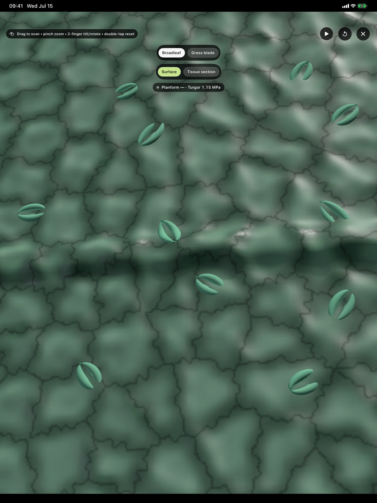
A synthetic broadleaf surface in the app’s immersive view.
04 Your work
Review one image or work through a batch. Keep the source, overlays, and tables together, then share the files through your device’s own tools.
Single-image exports include CSV instance tables, JSON summaries, the image, masks, and overlays, packaged for sharing.
Analysis runs on the device. No cloud inference or account is required. You choose when to export or share your work.
iPhone & iPad
Stomata analysis, on your device.